March 11, 2026
[THE IMPORTANCE OF EARLY DIAGNOSIS]
What types of glaucoma are there?
Primary open-angle glaucoma is the most prevalent type, with an unknown cause. It is distinct from secondary glaucoma, which results from corticosteroids, intraocular inflammation, tumors, trauma, or surgery.
In most cases, intraocular pressure (IOP) is elevated due to decreased drainage of aqueous humor (intraocular fluid in the anterior segment of the eye), compressing the optic nerve fibers. More rarely, IOP is normal, with the pathology associated with vascular lesions or optic nerve ischemia (more common in people suffering from vascular diseases, such as diabetes).
What are the symptoms?
Glaucoma usually manifests itself chronically and painlessly, with irreversible loss of visual field from the periphery to the center. As the visual field narrows, the patient may bump into objects and/or people from the side or suffer falls and accidents without realizing the cause. Without early diagnosis, the disease is only detected in advanced stages, culminating in tunnel vision or blindness.
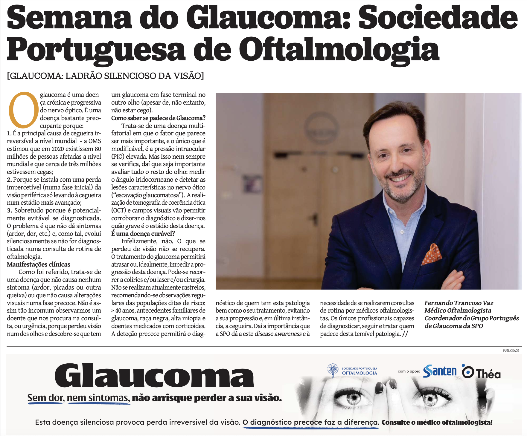
Glaucoma mainly manifests itself after the age of 40, and regular monitoring is essential, even if there are no symptoms.
In rarer cases, glaucoma manifests itself acutely, with symptoms that may include: eye pain, red eye, decreased vision, halos, headache. Of particular note are eyes with a narrower anterior chamber angle (where the aqueous humor drains), which suddenly "closes" in association with certain medications, abruptly hindering drainage (particularly in patients with anatomical predisposition, including an already narrow angle). Another example is acute IOP elevation associated with certain types of eye inflammation and infection.
EYE EXAMS ARE ESSENTIAL FROM THE AGE OF 40 ONWARDS AND FOR ALL PEOPLE WITH RISK FACTORS.
How to diagnose and monitor?
Regular eye exams are essential starting at age 40 and for all people with the following risk factors: family history of glaucoma, high IOP, black race, high myopia, and use of corticosteroids.
The initial consultation includes assessment of: visual acuity, IOP, anterior segment (biomicroscopy), and fundus, including the optic nerve (fundoscopy). In the event of changes, the following are performed: assessment of the anterior chamber angle (gonioscopy), measurement of corneal thickness, which can influence the measured IOP value (pachymetry), assessment of structural damage to the optic nerve (optical coherence tomography – OCT), and assessment of the visual field (perimetry). Follow-up is adjusted according to severity, including repeat examinations to assess the progression of the disease. //
Ana Sofia Lopes
Ophthalmologist
Portuguese Glaucoma Group of the SPO